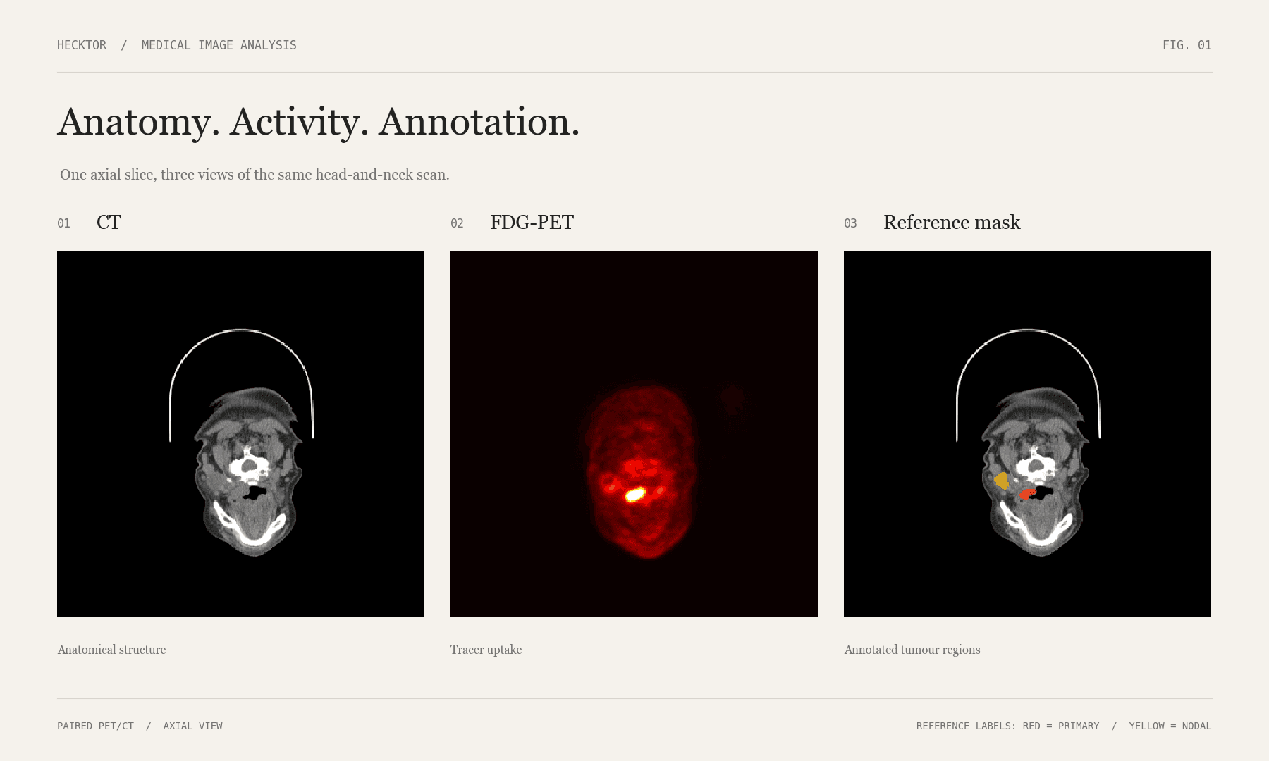
Can tumour segmentation support downstream cancer staging?
The project asked whether tumour segmentation from PET/CT imaging could provide useful information for downstream cancer staging. That meant solving two connected problems: first, producing a useful 3D tumour mask; second, turning that mask into structured features that could help predict T-stage and N-stage.
My contribution
- Contributed to PET/CT preprocessing and the tumour-segmentation workflow.
- Worked with tumour-burden and imaging-derived features.
- Used those features in downstream T-stage and N-stage experiments.
- Compared staging performance using balanced accuracy.
Problem & approach
At MIPSLAB, I contributed across the pipeline instead of working on one isolated model. I worked with PET/CT preprocessing, the 3D U-Net segmentation workflow, tumour-burden and imaging-derived features, and downstream staging experiments. The part that stuck with me was how connected everything was: a preprocessing choice could change the mask, the mask changed the features, and the features changed the staging result. The segmentation workflow uses PyTorch and MONAI for volumetric processing.
How it works
- 01
Prepare PET/CT
Prepare paired PET and CT volumes using volumetric medical-imaging preprocessing.
- 02
Segment tumours
Run a 3D U-Net-based segmentation pipeline to produce tumour masks.
- 03
Extract features
Extract tumour-burden and imaging-derived measurements from the masks.
- 04
Predict stage
Use those structured features in downstream T-stage and N-stage prediction experiments.
- 05
Evaluate
Evaluate segmentation with Dice and staging with balanced accuracy.
Data & evaluation
Data & inputs
Head-and-neck PET/CT imaging from the HECKTOR task. Segmentation and staging are connected through tumour-burden and imaging-derived features.
How it was evaluated
Project experiments reported approximately 0.7 segmentation Dice. T-stage balanced accuracy improved from about 0.312 to 0.585; N-stage improved from about 0.197 to 0.678. These are approximate project results tied to the experiment setup.
Results & progress
- ≈0.7
- Project segmentation Dice
- 0.312 → 0.585
- T-stage balanced accuracy
- 0.197 → 0.678
- N-stage balanced accuracy
Tumour masks and T/N-stage predictions
- Approximately 0.7 tumour-segmentation Dice.
- T-stage balanced accuracy improved from approximately 0.312 to 0.585.
- N-stage balanced accuracy improved from approximately 0.197 to 0.678.
Screenshots
Limits & lessons
Limitations
- These were research experiments under a specific project split and evaluation setup. They are not clinical deployment claims, and the pipeline would need independent validation before any clinical interpretation.
What I learned
- This project changed how I think about ML. It made it obvious that a useful system is not just dataset → model → metric. Preprocessing, feature design, evaluation, reproducibility, and understanding failure cases matter just as much as the model itself.
Next steps
- Compare different tumour-derived feature sets.
- Study failure cases instead of only aggregate metrics.
- Test how sensitive downstream staging is to segmentation quality.
- Validate the pipeline on independent data.